microdefectcv
MicroDefectCV
Adaptive OpenCV-based defect enhancement and segmentation for SEM and microstructure images.


![]()
![]()
A domain-specific computer vision toolkit for defect detection in perovskite solar cell SEM images. MicroDefectCV provides a reusable, mode-aware pipeline for pinhole and PbI₂ bright-particle detection that generalises to a wide range of microstructure images — no deep learning or labelled data required.
This package provides a lightweight classical computer vision baseline for defect enhancement and segmentation. It does not claim to replace deep learning methods on large annotated datasets.
Features
- 🔬 Six detection modes covering different perovskite morphologies and defect types
- 🧠 Auto mode that classifies image morphology from statistics alone
- 🧩 Grain boundary suppression for 3D and mixed-morphology images
- 📐 Needle crystal detection for elongated PbI₂ excess structures
- 📊 Defect statistics (count, area, area ratio) in one call
- 🖼️ Intermediate stage images for debugging and research
- ✅ Zero deep learning — pure OpenCV + NumPy, runs on CPU
- 📦 Pip-installable clean package structure
- 💻 CLI entry point — run
microdefectcvdirectly from any terminal after install
MicroDefectCV
MicroDefectCV is a computer vision toolkit for defect detection in perovskite solar cell SEM images.
PyPI
https://pypi.org/project/microdefectcv/
Installation
pip install microdefectcv
Or install from source:
git clone https://github.com/Sahilsonii/microdefectcv.git
cd microdefectcv
pip install -e .
Quick Start
import cv2
from microdefectcv import detect_defects

image = cv2.imread("sample_images/sem_image.png")

result = detect_defects(
    image,
    mode="auto",       # auto-selects morphology from image statistics
    min_area=20,
    return_intermediate=True
)

print(f"Defects found  : {result['defect_count']}")
print(f"Area ratio     : {result['defect_area_ratio']:.4f}")

mask     = result["mask"]         # binary defect mask
enhanced = result["enhanced"]     # CLAHE-enhanced image
contours = result["contours"]     # list of OpenCV contours
Detection Modes
| Mode | Target Defects | Image Morphology |
|---|---|---|
auto |
All | Auto-detected from statistics |
pbi2 |
PbI₂ bright particles + needles | Any |
pinhole |
Dark pinholes (small + large) | Any |
2d |
Both | 2D perovskite (flat morphology) |
3d |
Both + needles | 3D perovskite (grain suppression active) |
3d_2d |
Both + needles | Mixed 2D-3D morphology |
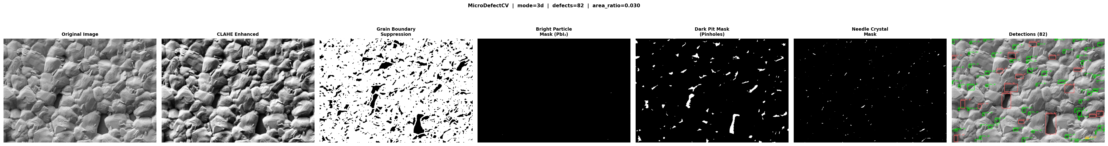
Method Pipeline
Input Image
    │
    ├─ Grayscale conversion (if BGR)
    ├─ SEM metadata bar removal
    ├─ Mode selection (auto or user-specified)
    ├─ Gaussian denoising + CLAHE
    │
    ├─ [3D / 3D-2D only] Grain boundary suppression mask
    │
    ├─ Bright particle detection (Top-Hat + dual percentile threshold)
    ├─ Dark pit detection       (Percentile threshold + micro-threshold)
    ├─ Needle crystal detection (Rectangular Top-Hat + aspect ratio filter)
    │
    ├─ Shape feature filtering (area, circularity, solidity, contrast)
    ├─ Non-maximum suppression (IoU-based)
    │
    └─ Output: mask, enhanced, contours, defect_count, defect_area_ratio
See docs/method_overview.md for full technical details.
Parameters
| Parameter | Type | Default | Description |
|---|---|---|---|
image |
np.ndarray |
— | Grayscale or BGR uint8 image |
mode |
str |
"auto" |
Detection mode (see table above) |
sensitivity |
float |
1.5 |
Sensitivity hint (reserved for tuning) |
min_area |
float |
20 |
Minimum defect area in pixels |
return_intermediate |
bool |
False |
Include per-stage pipeline images |
Quick Start Guide
Method 1: Command Line (Single Image)
After pip install microdefectcv, the microdefectcv command is available from any terminal — no need to navigate to a script folder.
# Auto-detect mode
microdefectcv "C:\Users\asus\Desktop\SEM annotation\3D perovskite with PbI2 excess\08-10.tif" --mode auto --min-area 20

# PbI2 bright particle + needle detection
microdefectcv "C:\Users\asus\Desktop\SEM annotation\3D perovskite with PbI2 excess\08-10.tif" --mode pbi2 --min-area 30

# Pinhole / dark void detection
microdefectcv "C:\Users\asus\Desktop\SEM annotation\3D perovskite with PbI2 excess\08-10.tif" --mode pinhole --min-area 20

# 2D perovskite (flat morphology)
microdefectcv "C:\Users\asus\Desktop\SEM annotation\3D perovskite with PbI2 excess\08-10.tif" --mode 2d --min-area 20

# 3D perovskite with grain boundary suppression
microdefectcv "C:\Users\asus\Desktop\SEM annotation\3D perovskite with PbI2 excess\08-10.tif" --mode 3d --min-area 20

# Mixed 2D-3D morphology
microdefectcv "C:\Users\asus\Desktop\SEM annotation\3D perovskite with PbI2 excess\08-10.tif" --mode 3d_2d --min-area 20
Method 2: Batch Processing (PowerShell)
Process an entire folder of images automatically:
Get-ChildItem -Path "path\to\folder" -Filter *.jpg | ForEach-Object {
    microdefectcv $_.FullName --mode auto
}
Method 3: Python API
Import and use directly in your own scripts:
import cv2
from microdefectcv import detect_defects
from microdefectcv.visualization import save_yolo_annotations

image = cv2.imread("path/to/image.jpg")
result = detect_defects(image, mode="auto", min_area=20)

print(f"Found {result['defect_count']} defects!")
save_yolo_annotations(result["detections"], image.shape, "outputs/labels.txt")
Results
Comparison
| Method | Suitability | Notes |
|---|---|---|
| Global Threshold | Low | Fails under uneven SEM lighting |
| Otsu | Low–Medium | No domain adaptation |
| CLAHE + Otsu | Medium | Better contrast, still single-class |
| Canny | Edge-only | Not suitable for void/particle detection |
| MicroDefectCV | High | Adaptive, mode-aware, domain-specific |
Running Tests
pytest
Use Cases
- Perovskite solar-cell SEM — pinhole and PbI₂ crystal detection
- Thin-film defect inspection — dark voids and bright particle segmentation
- Microstructure void detection — general SEM / optical microscopy
- Coating and surface QC — surface dark defect segmentation
- Classical CV baseline — compare against DL models on annotated datasets
Citation
If you use MicroDefectCV in academic work, please cite:
@software{microdefectcv2025,
  title  = {MicroDefectCV: Adaptive OpenCV-based Defect Segmentation for SEM Images},
  author = {Sahil Soni},
  year   = {2025},
  url    = {https://github.com/Sahilsonii/microdefectcv}
}
Roadmap
- Annotated SEM benchmark dataset
scripts/evaluate.pyevaluation script- Hyperparameter search / sensitivity analysis
- Optional integration with OpenCV-contrib
License
MIT — see LICENSE.